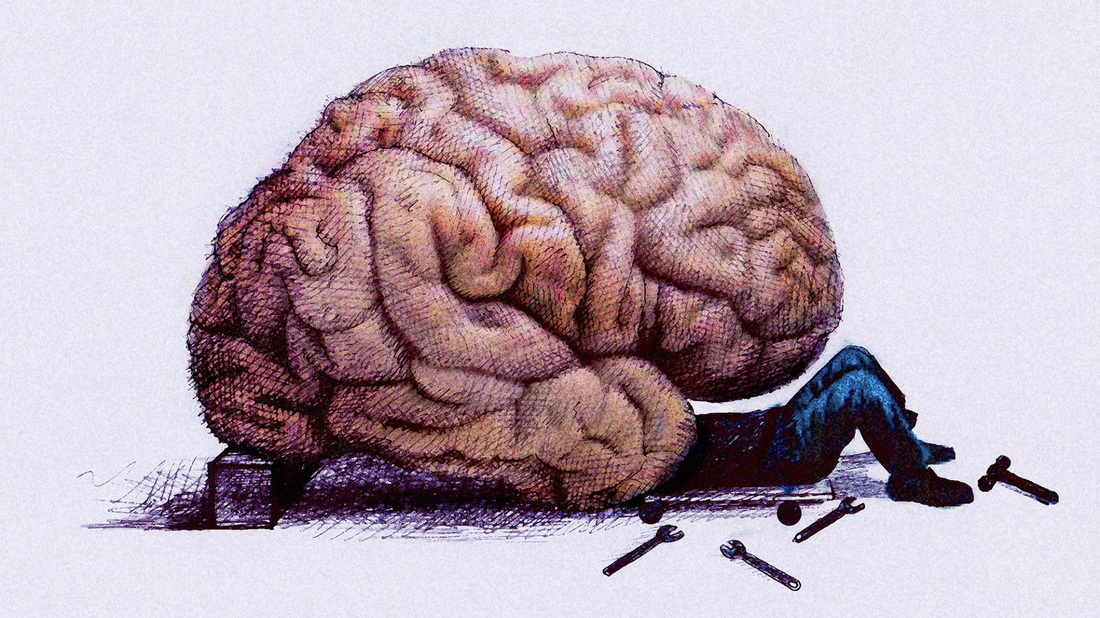
LATERALIZATION AND PLASTICITY OF THE BRAIN
The lateralization of brain function refers to how some neural functions, or cognitive processes tend to be more dominant in one hemisphere than the other. The medial longitudinal fissure separates the human brain into two distinct cerebral hemispheres, connected by the corpus callosum. Although the macrostructure of the two hemispheres appears to be almost identical, different composition of neuronal networks allows for specialized function that is different in each hemisphere.
Better understanding how lateralization relates to brain function is relevant to many people, among them: academic researchers, medical clinicians, neurological patients, educators and left-handers. Clarifying the relationship between handedness and functional brain specializations, and learning more about the developmental and neurobiological mechanisms that underlie these relationships, may help us better understand a wide range of seemingly unrelated issues such as dyslexia, stuttering, human variation, comparative brain research, developmental neurobiology of the brain, and the origins of human language.
Brain plasticity (from the Greek word ‘plastos’ meaning molded) refers to the extraordinary ability of the brain to modify its own structure and function following changes within the body or in the external environment. The large outer layer of the brain, known as the cortex is especially able to make such modifications.
Brain plasticity underlies normal brain function such as our ability to learn and modify our behavior. It is strongest during childhood — explaining the fast learning abilities of kids — but remains a fundamental and significant lifelong property of the brain. Adult brain plasticity has been clearly implicated as a means for recovery from sensory-motor deprivation, peripheral injury, and brain injury. It has also been implicated in alleviating chronic pain and the development of the ability to use prosthetic devices such as robotic arms for paraplegics, or artificial hearing and seeing devices for the deaf and blind.
NEUROSENSORY ORGANIZATION
OF SENSATION AND PERCEPTION
FOR COMMUNICATION
Recognition and identification of a sensory stimulus is a critical function and affects the evaluation of communication skills. Perception of a word requires adequate hearing acuity, but the stimulus must be recognized as a word. The features must be visibly, audibly, or tactically perceived before an object can be recognized.
Sight
We communicate messages through sight by using visual signals that include facial expressions, gestures and posture (or body language). We receive these signals by using our sense of sight.
When we look at something, light bounces off the object and onto the pupil in the eye. The light crosses the lens of the eye, the picture becomes focused, and then turns upside down. The picture then shines on the retina, at the back of the eye. A retina contains rod cells and cone cells, which are both photoreceptors. These cells let your eye see colours and details. The optic nerve sends a message of this picture to your brain, where the picture is turned the right way up. Your brain then tells you what response you should make to the object that you can see.
Hearing
The most common way for humans to communicate is by the sound made through speech. One person speaks and the other person receives the message by hearing it with their ears.
The ear has three parts: the outer ear, the middle ear, and the inner ear. Sounds reach the outer ear first, then travel into the ear canal and finally reach the eardrum. The eardrum is a thin piece of tissue that separates the outer ear and the middle ear. There are three tiny bones in the middle ear that make sounds louder. Sounds from the middle ear travel to the inner ear, where they make tiny hairs inside the cochlea (which looks like a snail) move around. The receptor cells then send signals along the auditory nerve to the brain. The brain changes these signals back into meaningful sound that we can understand.
Taste
We can communicate by receiving messages through taste. Babies make good use of communicating with their world by tasting things around them.
Taste lets you enjoy the flavour of your favourite foods. You can tell if food has gone off because it tastes unpleasant. Taste also tells you if something is dangerous or poisonous, although you should never taste anything if you think that it might be unsafe. If you look carefully at your tongue you will see tiny little bumps all over it - these are called taste buds. There are four different types of taste buds on your tongue. At the front of your tongue you can taste sweet, on both sides of the tongue you taste sour, at the back you taste bitter, and all over your tongue you taste salty.
Touch
We communicate with touch by feeling things. People hug to show that they are happy to see each other, shake hands to show that they agree, or put their arms around a person who is upset.
We feel messages that are communicated to us by touch through touch receptors. These are located in groups around the skin and look a bit like tiny onions. When they are squeezed, the layers rub against each other and send electrical signals to the brain. Some touch receptors are more sensitive than others. Sensitive touch receptors can be found on different parts of your body, including your face and your fingers.
Smell
We send and receive messages through smell. We can smell dangerous things like smoke from a fire or poisonous gas. We can also smell pleasant things like flowers or a freshly baked cake. Smell communicates powerful messages to our noses.
When we breathe, air goes into the nose through the nostrils. The air then travels down the back of the mouth and into the throat. Any smell, or odour, that passes through the nasal cavity is stuck to the mucus in your nose. The tiny hairs in your nose, called sensory hairs, sense the odour and send messages to your brain where the smell is identified. The smell receptor cell, which responds to the chemicals in the mucus in your nose, is positioned high up behind the nose.
NEUROMOTOR AREAS THAT
ARE IMPORTANT FOR
SPEECH AND COMMUNICATION
Pyramidal Tracts
The pyramidal tracts include both the corticospinal and corticobulbar tracts. These are aggregations of upper motor neuron nerve fibres that travel from the cerebral cortex and terminate either in the brainstem (corticobulbar) or spinal cord (corticospinal) and are involved in control of motor functions of the body.
The corticobulbar tract conducts impulses from the brain to the cranial nerves. These nerves control the muscles of the face and neck and are involved in facial expression, mastication, swallowing, and other functions.
Extrapyramidal System (and Tracts)
The extrapyramidal system is a biological neural network that is part of the motor system causing involuntary movements. The system is called "extrapyramidal" to distinguish it from the tracts of the motor cortex that reach their targets by traveling through the "pyramids" of the medulla. The pyramidal pathways (corticospinal and some corticobulbar tracts) may directly innervate motor neurons of the spinal cord or brainstem (anterior (ventral) horn cells or certain cranial nerve nuclei), whereas the extrapyramidal system centers on the modulation and regulation (indirect control) of anterior (ventral) horn cells.
Extrapyramidal tracts are chiefly found in the reticular formation of the pons and medulla, and target neurons in the spinal cord involved in reflexes, locomotion, complex movements, and postural control. These tracts are in turn modulated by various parts of the central nervous system, including the nigrostriatal pathway, the basal ganglia, the cerebellum, the vestibular nuclei, and different sensory areas of the cerebral cortex. All of these regulatory components can be considered part of the extrapyramidal system, in that they modulate motor activity without directly innervating motor neurons.
Cortico-Cerebellar System
The cerebellum can influence a variety of motor control centers in the brain, including the motor cortex, through its output pathways. Inputs to the cerebellum provide it with access to sensory information that serves as feedback to guide learning. Computational models of its circuitry, and evidence from neurophysiological studies, show that cerebellar circuits are capable of plasticity that could support the storage of ‘motor memory’. Our work uses functional MRI to investigate the cerebellar mechanisms of motor skill in the human brain by studying its responses to error feedback and charting the dynamics of its activity while people acquire motor skills in the scanner.
EXECUTIVE FUNCTIONS AND LEARNING

Executive function’s relationship to learning is that it helps us to focus more on the task at hand which helps us to be more efficient in absorbing and processing the information. It makes us flexible of how we think. It is aslo a way of evaluating ourselves if how do we comprehend things appropriately and lastly it also helps us to organize our thoughts and keep track of our train of thoughts.
Structures:
1. Limbic association area
-Located in the anterior-ventral portion of the temporal lobe (parahypocampal gyrus)
- It links emotion with many sensory inputs.
- Important in learning and memory.
(olfactory bulb, hypocampus, hypothalamus, amygdala, anterior thalamic nuclei, fornix, mammilliary body, septum pellucidum, cingulate gyrus, parahypocampal gyrus, limbic cortex)
2. Posterior association are:
- Located at the junction of occipital, temporal and parietal lobes.
- It links information from primary and unimodal sensory areas.
- Important in perception and language.
- This is what gives us our spatial awareness of our body. It is the kinesthetic sense that is very strong in professional dancers or athletes for example.
- Anterior association area
- Located in the prefrontal cortex.
-It links information from other association areas.
- Important in memory, planning, and higher-order concept formation.
- It has a lot to do with thinking and making judgments and it’s where you understand what’s socially acceptable, how to behave; just anything related to heavy human conditioning.
